Chromosome
If you look at the onion root under a microscope, you can see cells of different shapes and sizes, such as cells with an apparent nucleus, cells that appear to be split into two cells, and cells that have no nucleus and that have structures that look like short strings or rods.
Structures that look like short strings or rods in cells are called chromosomes, which appear when cell division occurs. Therefore, by observing the shape and movement of chromosomes, you can distinguish the process of cell division.
Chromosomes are loosened like thin threads when cells don’t divide (DNA). Before the cells divide, the DNA is replicated and thick, shortened together. In Prophase and Metaphase, one chromosome consists of two strands called chromatid, each with the same genetic information. (Replicated)
Mitosis
| Interphase | 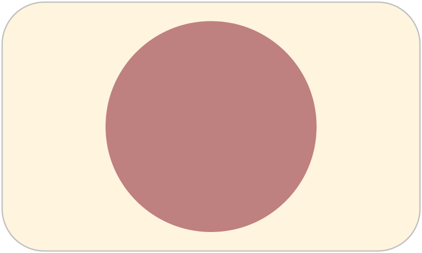 |
In preparation for cell division, the gene is replicated and its amount doubles. The nuclear membrane is visible. |
|---|---|---|
| Prophase | 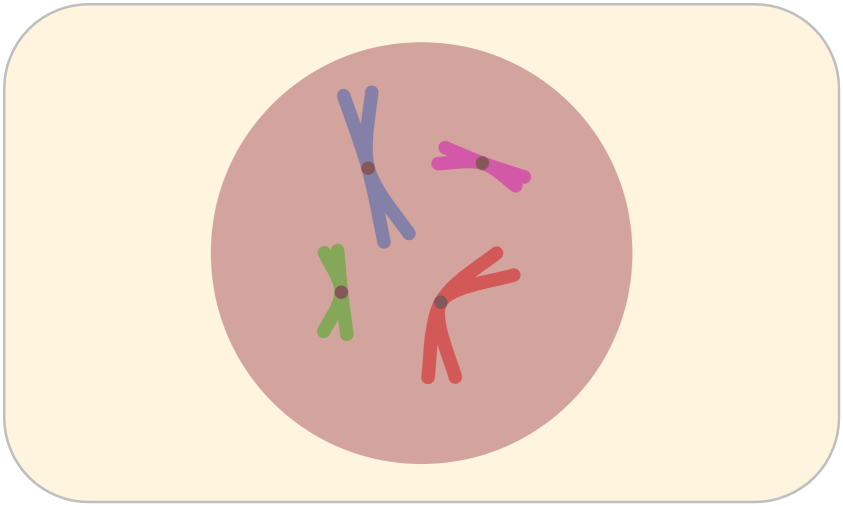 |
The chromosome appears as two strands of chromatid. The nuclear membrane disappears and spindles are formed. (The simulation doesn’t show the spindle.) |
| Metaphase | 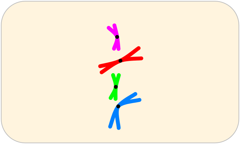 |
Chromosomes are arranged in the center of the cell. |
| Anaphase | 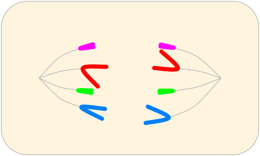 |
Each chromosome is separated by spindles and moved to the anode. |
| Telophase | 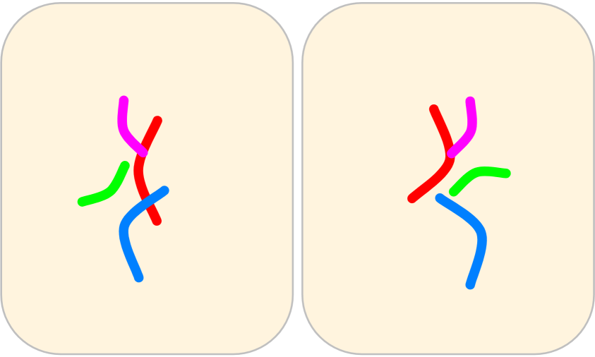 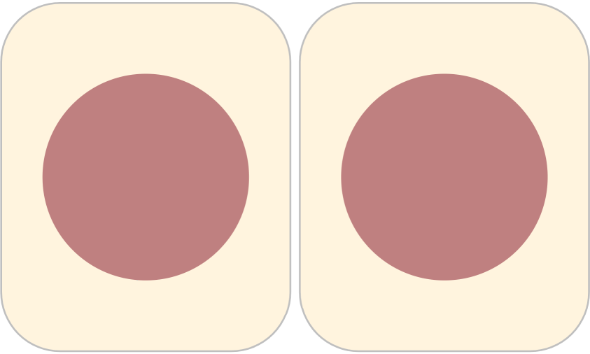 |
The condensed chromosome is released, the nuclear membrane is formed, and the nucleus is rebuilt. Two daughter cells are then formed. |